
Standards and 3D Printing Human Tissue
Fifty-three years ago, the Bee Gees posed a question: “How do you mend a broken heart?” At the time, few would have predicted that this metaphorical query might someday be answered with “collagen, hyaluronan, and a 3D printer.”
Although researchers are still years away from being able to produce complex organs like livers, kidneys, and yes, hearts, advancements in additive manufacturing (AM) technology — often referred to as 3D printing — have transformed the idea of fashioning organic tissue into anatomical replacement parts from far-fetched concept to very real and potentially revolutionary reality.
Adaptation of specific AM processes to the creation of what are known as tissue engineered medical products (TEMPs) is still in its early stages. But AM technology, which builds three-dimensional objects by adding successive layers of material as directed by computerized design files, is no stranger to medical applications. For some orthopedic TEMPs, the material of choice is titanium — a strong, lightweight metal that offers excellent biocompatibility.
FOR YOU: The Future of Additive Manufacturing Standards
As AM continues to evolve into a viable option for addressing medical issues, so too must material specifications, test methods, and other standards. Members of ASTM International’sl committees on medical and surgical materials and devices (F04) and additive manufacturing technologies (F42) are hard at work on a variety of standards covering both tissue-based and polymeric or metallic AM feedstocks.
Heart Valves
One of the most dramatic potential uses for AM in a healthcare context is the fabrication of replacement heart valves to treat both congenital defects and degenerative disease. A new standard guide designed to support ongoing efforts to refine and improve these printed valves is approaching the finish line, with final approval expected this summer.
Sandy Williams, Ph.D., is founder and president of Access Biomedical Solutions. An active ASTM member since 2006 and the technical contact for the new standard guide for heart valve TEMPs (WK84054), she defines TEMPs as “essentially medical products that repair, modify, or regenerate a patient’s cells, tissues, and organs.”
“Additive manufacturing methods are sometimes utilized to develop TEMP heart valves,” Williams says. “3D printing is used with biological and synthetic biomaterial scaffolds, and bioprinting introduces cells to the 3D constructs. The new standard guide is intended to fill in the gap for how TEMP heart valve manufacturers could characterize and assess their products in vitro,” incorporating test protocols designed to assess biochemical, mechanical, biological, and other properties of these products.
One of the challenges of putting the guide together is the fact that TEMP heart valves are still in a relatively early stage of development and are not yet widely used. “Manufacturers have had to work closely with regulatory bodies such as the U.S. Food and Drug Administration [FDA] to determine the best ways of assessing TEMP safety and efficacy based on each specific valve technology,” explains Williams. She gathered valuable input from several of these manufacturers for inclusion in the standard guide.
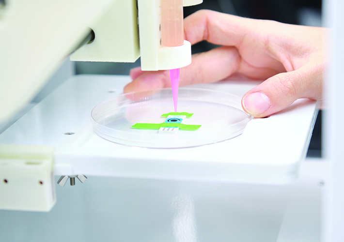
Bioinks and Bioprinting
Examination of another new standard guide nearing completion provides an opportunity to gain a better understanding of what the terms “scaffold” and “bioprinting” mean.
The standard guide for bioinks used in bioprinting (WK74668) “defines key terms to provide a common language for all stakeholders,” according to Carl Simon, Jr., Ph.D., a biologist in the Biosystems and Biomaterials Division of the National Institute of Standards and Technology (NIST) and F04 member. “It describes many issues that must be considered for successful bioprinting, such as sterility, bioink biocompatibility, printing parameters, and strategies to keep cells alive during printing.”
Broadly speaking, bioink is a material used for bioprinting TEMPs for biomedical applications. “Bioprinting is the process of making devices for biomedical applications using AM,” says Simon. Typically, cells would be suspended in the bioink and printed onto a scaffold, layer by layer, using a bioprinter.
Bioink is usually a hydrogel, a hydrophilic polymer that absorbs water to make a clear, jelly-like material. The polymer can be naturally derived (collagen or hyaluronan are the usual base components) or made up of degradable synthetics. It starts out soft so it can be extruded and is then hardened by a physical mechanism (curing, polymerization, phase transition) after printing so the end result maintains its intended shape.
A bioink is often used to 3D-print a scaffold, the substrate upon which cells may grow and regenerate tissue. A scaffold is generally three-dimensional and porous, to provide space for cells and nutrients to flow and adhere, generating tissue. The bioink may contain cells during the bioprinting process so that the final construct is already fully loaded with cells. Other scaffold-building processes include salt leaching, phase separation, and electrospinning. At this time, only a fraction of scaffolds are created via AM.
The day when bioprinting human tissue is commonplace is fast approaching.
“In the context of the new ASTM standard guide, the bioink is the material for the scaffold,” Simon notes. “One may print the bioink into a scaffold and seed cells after printing, or cells can be mixed within the bioink and then printed into a porous scaffold structure.”
As with most new standards, defining the scope for this guide required a lot of give and take in the five-plus years since work began. “There are many opinions about what constitutes bioprinting,” says Simon. “If you include all of them, the standard becomes so broad and dilute that it has little value.”
The team decided to focus on extrusion-based bioprinting because it is the most advanced AM process in terms of medical applications. Simon describes extrusion printing as: “Like squeezing liquid out of syringe. It’s done in automated fashion using a piston with a screw mechanism or pneumatically with air pressure.”
The standard guide also addresses other bioprinting methods, including electrospinning, electrospray, droplet-based, inkjet-based, and laser-assisted.
Regenerating Muscle Tissue
The creation of complete, fully functioning organs via AM is no longer a science fiction fantasy, though it will undoubtedly take years of research and experimentation to get from here to there. However, the use of AM to regenerate muscle tissue is much further along, and another standard currently in development will help support additional progress in this area.
Volumetric muscle loss can occur in different ways. Blast injuries, tumor resection, and compartment syndrome (excessive pressure build-up inside an enclosed muscle space in the body caused by bleeding and swelling, often after a limb is broken) are some of the most common causes. Less severe conditions like a torn rotator cuff in the shoulder or a ruptured muscle are also candidates for this type of treatment, according to Michael McClure, Ph.D., assistant professor in the Department of Biomedical Engineering at Virginia Commonwealth University and F04 member.
McClure is a member of the team working on the new standard guide for pre-clinical testing considerations for materials used to regenerate muscle following volumetric muscle loss injuries (WK78974). He explains that “The ultimate goal is to create new tissue in a compromised area,” and that the tissue itself is specific to the situation it is being produced to address: muscle, heart, heart valve, tendon, ligament, bone, skin, etc.
This work item focuses specifically on muscle tissue and the materials used to bioprint it. “Primarily natural polymers are used,” McClure says. “Collagen, hyaluronic acid, decellularized muscle, decellularized bladder, decellularized small intestine submucosa. Each product can vary in its elastic modulus and composition, which can affect the regenerative capacity of the material.”
The Cambridge Dictionary defines decellularization as “a process in which cells are removed from the tissue surrounding them.” The resulting extracellular matrix (ECM) becomes the biologic scaffold upon which a desired TEMP can be constructed.
“The standard guide is designed to provide information to researchers about different considerations when designing a treatment for volumetric muscle loss. These considerations can include such areas as wound location, muscle-fiber types, and muscle architecture,” says McClure.
Why Titanium?
It should come as no surprise that titanium is in the middle of the standards-development activity surrounding the use of AM in medical applications. For decades this strong, lightweight metal has had one foot in the operating room and the other in the AM laboratory.
There are two primary reasons titanium has been used for surgical implants since the 1950s. The first is its ability to interact in a nonreactive way with human tissue — in other words, biocompatibility. This trait can be attributed to passivation, the process by which the metal forms an oxide layer on its outer surface that prevents it from reacting with tissue it comes in contact with.
The second is osseointegration, which is defined as the creation of a structural and functional connection between living bone tissue and the surface of load-bearing implants such as hip cups, prosthetics, and spinal-fusion devices.
READ MORE: The 5 Most Important Additive Manufacturing Standards
Titanium has also proven to be a good fit for additive manufacturing. In powder form, the metal and its alloys are used as feedstocks in several AM processes, including laser powder bed fusion (LPBF), electron beam melting, and directed energy deposition. Parts made in this way deliver benefits in terms of strength-to-weight ratio, corrosion resistance, and other properties in industries like aerospace, automotive, and chemical.
Medical is another such industry. “The LPBF process is optimum for consolidating the titanium powder into the required shapes for orthopedic products,” notes Rod McMillan, engineering fellow at DePuy Synthes, a Johnson and Johnson MedTech company, and a member of both F42 and F04. “There is a good fit between the dimensions required and the spot size of the laser. Titanium welds well and the laser fusion process has been developed in recent years to provide excellent material properties.”
Matthew Di Prima, Ph.D., materials scientist in the FDA’s Division of Applied Mechanics who also participates in F42 and F04, adds that “Since LPBF is dominant in medical applications, our subcommittee has primarily focused on this AM technology.” That focus is currently directed at modifying existing standards that were not optimized for medical purposes at the time of their original publication.
New Material Specifications
Two grades of titanium are commonly used in AM applications: grade 5 (Ti-6Al-4V) and grade 23 (Ti-6Al-4V ELI). Thanks to lower oxygen content that slightly reduces its static strength, grade 23 delivers improved fatigue performance compared to grade 5, but each has been utilized for implantable devices for many years, and each is covered by a standard specification: the specification for additive manufacturing titanium-6 aluminum-4 vanadium with powder bed fusion (F2924); and the specification for additive manufacturing titanium-6 aluminum-4 vanadium ELI (extra low interstitial) with powder bed fusion (F3001), respectively.
Because these specifications were designed for general AM applications, members of the F42 medical/biological subcommittee (F42.07.03) decided to create medical application-specific versions. The grade 23 standard (F3001) is the first to be modified, with plans to address the grade 5 standard in the future.
“The chemistry and material requirements are generally the same as in F3001,” Di Prima says. “However, we are proposing consolidating the classes in F3001 to include only those relevant to medical applications, updating the heat-treatment language, and including language on medical validation. The committee hopes this will make the standard more useful for medical-device companies and regulatory bodies.”
McMillan points out that the new specification will define the chemistry necessary for AM medical devices, and that: “It will also define the controls necessary to produce a high-quality part in the AM process that meets the needs for long-term implantation in humans.”
F3001 is also the starting point for the other new titanium/LPBF standard currently under development. This one will require a new number as the suggested change involves replacement of the alloying elements, switching out aluminum and vanadium (Ti-6Al-4V) for niobium and zirconium (TiNbZr).
“While this new specification will also be for the PBF process, it’s likely that there are device applications where the mechanical properties of titanium with niobium and zirconium would make it more attractive than titanium with aluminum and vanadium,” Di Prima says. “This specification is about providing more options for medical device manufacturers when selecting an AM material. Our hope is that once we release these first medical AM material specifications, we can quickly release several more covering other materials of interest to the medical AM community.”
More in the Pipeline
There is clearly a lot of ongoing work in the “medical wing” of the AM universe. Some of this work is described here, but there are other initiatives under way that deserve a mention.
For example, the members of F42.07.03 are working on two new test methods relevant to medical applications of AM. One involves assessing the presence of residual powder in medical devices produced using PBF. “Any residual powder in a device could potentially migrate out of the device or inhibit tissue growth,” Di Prima explains.
“Metal powder from the printing process is a new type of contaminant,” says McMillan. “And it is necessary to develop techniques to assess the efficacy of the cleaning process to remove it.”
The other new test method is based on a gravimetric approach to determining the density of 3D-printed lattice structures. Because these structures are incorporated into implants to promote boney in-growth, it is important to ensure that pore size and the size of the lattice struts are correct. “This test also provides the opportunity to indirectly measure the stability of the printing process,” McMillan says.
Test methods will also be the focus of the upcoming virtual event, “AM Medical Device Verification Workshop” scheduled for March 19, which will explore build verification approaches other than the kind of tensile testing delineated in standard test methods for tension testing of metallic materials (E8).
Di Prima provides a concise summary of the ultimate objective of all this activity: “AM has been successfully adopted by the medical device industry, and with F42.07.03 we are hoping to develop standards that will address the needs of the medical community and reduce the barrier of using AM technology in this space.” ■
More on Tissue-Engineered Medical Product (TEMPs)
TEMPs are classified using a wide range of materials. The end product is used to repair, modify, or regenerate a recipient’s cells, tissues, and organs or their structure and function. TEMPs can be developed to be “off the shelf” or can be created at a patient’s bedside or within the patient. This involves the use of degradable synthetic polymers, natural polymers like collagen or hyaluronan, and cells. The goal is to regenerate new tissue in a compromised area, but the tissue itself is only specific for the application it is being used to treat. It could be muscle, heart, heart valve, tendon, ligament, bone, skin, and more.
When it comes to additive manufacturing (AM), synthetic and natural polymers can be used to create AM TEMPs. The primary base components are collagen or hyaluronic acid. Others will use crosslinkers to help stabilize the gel such as methacrylates, which assists with better control over AM. During the process of making the TEMP cells, it can be added while printing or after printing is complete. This is very different from metals, because the engineer needs to pay close attention to the gelation time, spread of the gel, whether it retains its shape, and consistency from batch to batch. Small changes in chemistry could yield large variations.
READ MORE about the AM Medical Device Verification Workshop.
Jack Maxwell is a freelance writer based in Westmont, N.J.
 SN Home
 Archive
 Advertisers
 Masthead
 RateCard
 Subscribe
 Email Editor